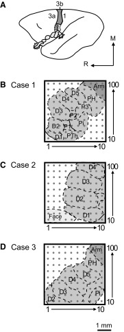
Fig. 2.
Schematic reconstructions show 100-electrode array placement in area 3b of 3 owl monkey cases. A: a schematic of the owl monkey brain is shown with the area 3b body representations highlighted as area 3a and area 1 neurons tended not to respond well to light tactile stimulation under the anesthetic conditions of these experiments. Gray shading indicates the area 3b hand representation. Light gray shading indicates the area 3b face and oral cavity representations, portions of which are hidden from the cortical surface, but are revealed by “unfolding” the schematic representation. Dark gray indicates the rest of 3b. B–D: electrode locations in each owl monkey case were approximated based on examination of myelin-stained sections of flattened cortex in which area 3b stains more darkly than surrounding areas, with a myelin-poor septum indicating the border of the hand and face representation. Occasionally, myelin-poor septa can seen between digit representations in flattened cortex preparations, but in our cases, we used the results of receptive field mapping during recording experiments to estimate the digit and palm pad representations. The placements of the 4 × 4 mm array in each case tended to cover a large part of the hand representation. The 1 mm scale bar corresponds to the size of the array schematics for all 3 owl monkey cases.

