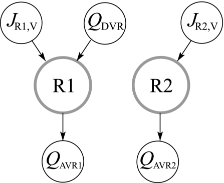
Fig. 3.
Steady-state blood flow patterns at a given medullary level. QDVR, DVR blood source; JR1,V and JR2,V, net transtubular water fluxes from tubules and vasa recta into R1 and R2, respectively; QAVR1, QAVR2, net fluid accumulation drained by AVR1 and AVR2 from R1 and R2, respectively. Arrows indicate flow direction.

