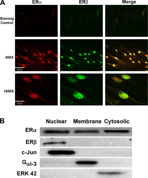
Fig. 2.
Localization of ERs in ASM cells. A: immunocytochemical staining of enzymatically dissociated ASM cells from female patients followed by confocal imaging demonstrated expression of both ERα (Cy3-conjugated secondary antibody; red) and ERβ (Alexa Fluor 488-conjugated secondary antibody; green). Top shows secondary staining controls, whereas middle and bottom shows ERα and ERβ expression (left, middle) as well as colocalization (merged images with yellow) at 2 different magnifications. ERα and ERβ are present at the plasma membrane as well as in the cytosol and nucleus, albeit to different extents. B: ER expression was determined in whole ASM cell lysates separated into heavy (nuclear/Golgi), membrane, and cytosolic fractions. Complementary to immunocytochemical staining, ERα was expressed by all 3 fractions, whereas ERβ expression was limited to the heavy fraction. Purity of the cell fractions was tested by blotting for proteins exclusive to each fraction. B represents original scan contrasts and brightness, which differed between blots.

