
Figure 2.
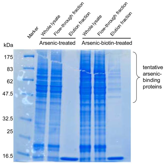
SDS-PAGE and Coomassie blue staining of the proteins from MCF-7 cell lysate. Three fractions were loaded, whole lysate, flow-through fraction which was unbound by streptavidin, and elution faction which was pulled down by streptavidin.
Official websites use .gov
A
.gov website belongs to an official
government organization in the United States.
Secure .gov websites use HTTPS
A lock (
) or https:// means you've safely
connected to the .gov website. Share sensitive
information only on official, secure websites.
SDS-PAGE and Coomassie blue staining of the proteins from MCF-7 cell lysate. Three fractions were loaded, whole lysate, flow-through fraction which was unbound by streptavidin, and elution faction which was pulled down by streptavidin.