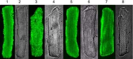
Fig. 1.
Intracellular localization of endogenous p21-activated kinase-1 (Pak1) regulated by bradykinin (BK) in rat ventricle myocytes. Lanes 1, 3, 5, and 7: confocal images with immunofluorescence signals for Pak1. Lanes 2, 4, 6, and 8: transmitted light images of the myocytes. Lanes 1 and 2: control myocytes without BK treatment. Lanes 3 and 4: myocytes treated with 0.1 μM BK. Lanes 5 and 6: myocytes treated with 1 μM BK. Lanes 7 and 8: myocytes treated with 10 μM BK. For immunofluorescence, the primary antibody was from Santa Cruz (sc-881). The secondary antibody was FITC-conjugated anti-rabbit IgG from Sigma (F-9887).

