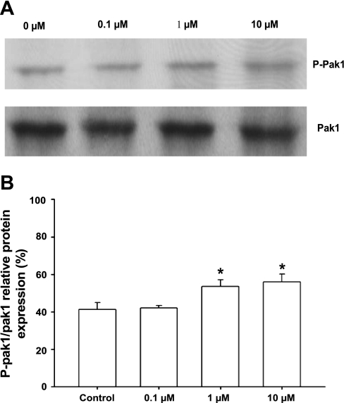
Fig. 2.
A: Western blot analysis of autophosphorylation of endogenous Pak1 in myocytes treated with BK at different concentrations. A, lanes 1–4: myocytes treated with BK at 0, 0.1, 1, and 10 μM, respectively, for 10 min. Pak1 autophosphorylation was detected by phospho (p)-specific (Pak1 Thr423) antibody. Total Pak1 served as an internal control. B: histogram showing increase of Pak1 Thr423 autophosphorylation normalized to total Pak1. Results shown are representative of 3 independent experiments. p-Pak1 Thr423 levels are expressed as percentage of total Pak1. *P < 0.05 compared with control without BK exposure.

