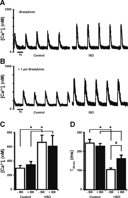
Fig. 6.
Intracellular Ca2+ concentration ([Ca2+]i) transients recorded from representative field-stimulated single adult rat ventricular myocytes in control conditions and after application of 100 nM Iso in an untreated cell (A) and a cell pretreated for 20 min with 1 μM BK (B) are shown. Summaries of peak [Ca2+]i transients (C) and the [Ca2+]i-transient time constant of decay (τdecay; D) from the same set of records are shown. Data are presented as means ± SE. *P < 0.05 compared with control; #P < 0.05 compared with −BK.

