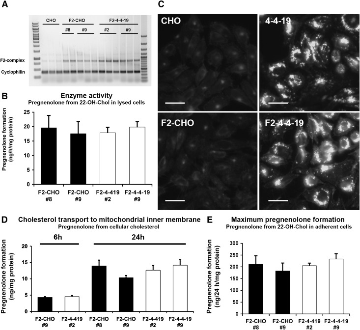
Fig. 2.
F2-CHO and F2-4-4-19 cells grown in the presence of serum transport the same amount of cholesterol to the mitochondrial inner membrane. A: F2-complex and cyclophilin mRNA in four monoclonal CHO cell lines stably expressing F2 and in parental CHO cells measured by RT-PCR, with each cell line in triplicate (clone #8 in duplicate). Lane 1, 1 kB ladder; lane 16, 100 bp ladder. B: CYP11A1 enzymatic activity in cell lysates of the four F2 clones shown as pregnenolone production from 22-OH-Chol in the presence of detergent and an NADPH regenerating system. Data are means ± SEM of four independent experiments in duplicate. C: Representative CHO, F2-CHO, 4-4-19, and F2-4-4-19 cells were fixed and stained with filipin to visualize unesterified cholesterol. Bars = 40 µm. D: Pregnenolone formation from endogenous cholesterol sources during 6 or 24 h in intact, adherent F2-CHO and F2-4-4-19 cells. E: Maximum rate of pregnenolone formation from excess membrane-permeable precursor 22-OH-Chol during 24 h in intact, adherent F2-CHO and F2-4-4-19 cells. In D and E, data are means ± SEM, three independent experiments in triplicate.

