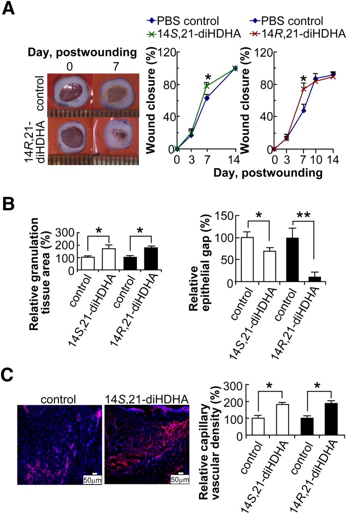
Fig. 3.
14S,21-diHDHAs and 14R,21-diHDHAs promote wound healing. Splinted excision wounding was conducted on Balb/c mice as in Fig. 1, followed by administration of 14S,21-diHDHA or 14R,21-diHDHA to wounds as detailed in “Materials and Methods.” Treatment with vehicle PBS saline was the control. Results are mean ± SEM, n = 5, *p < 0.05 and **p < 0.01 as compared with the control. A: 14,21-diHDHA accelerate wound closure. Representative photographs of wounds at days 0 and 7 postwounding show wound closure (left). Wound closure (%) was measured for the treatment with 14S,21-diHDHA (middle) and 14R,21-diHDHA (right). B: 14S,21-diHDHA and 14R,21-diHDHA promotes granulation tissue formation and reepithelialization in wounds by increasing the total granulation tissue area (left) and reducing epithelial gap (right), as determined in hematoxylin-eosin stained cryosections of wounds in comparison with PBS control. C: 14S,21-diHDHA and 14R,21-diHDHA increase the capillary vascular density in wound beds. Micrographs (left) show vasculatures as CD31+ cells in wound cryosections stained with rat anti-mouse CD31 antibody then with PE-goat anti-rat IgG (red); nuclei were stained with Hoechst 33342 (blue). Capillary vascular densities were quantified (right) as CD31+ cells in wound-bed/field relative to PBS control. Cryosections were conducted on wound skin collected immediately after sacrifice of the mice at day 4 postwounding.

