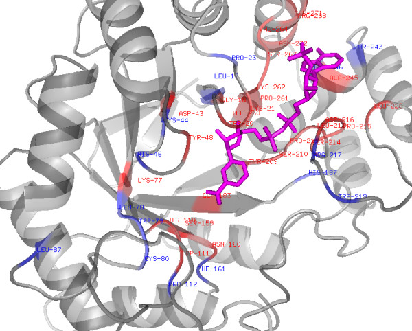
Figure 2.

Structure of human Aldose reductase (2ACS) showing prediction of NAD interacting residues by NADbinder. NAD shown in magenta, True positives in red and False positives in blue colour (only the portion of protein with residue mentioned is shown here).
