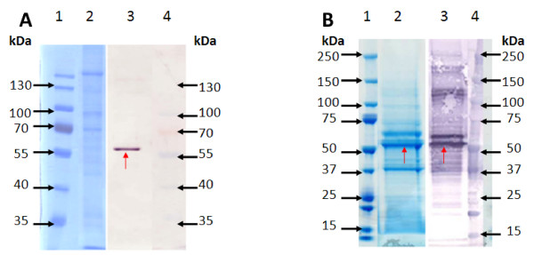
Figure 5.
Comparison of PB and BV VLP banding pattern differences as visualised on a Coomassie stained gel and western blot. Coomassie Blue stained 10% denaturing polyacrylamide gels are shown on the left of 5A and 5B and anti-p24 stained western blots are shown on the right of 5A and 5B. Protein size markers on gels and blots are indicated in kDA. Red arrows indicate the Gag Pr55 band. A- Purified PB VLPs. Lanes 1 and 4 - MW marker. Lane 2- Coomassie Blue stained PB VLP preparation (30 ul of purified sample, corresponding to 6 pg p24). Lane 3- PB VLP preparation detected with anti-p24 antibody (30 ul of purified sample, corresponding to 6 pg p24). B- Purified BV VLPs. Lanes 1 and 4 - MW marker. Lane 2- Coomassie Blue stained BV VLP preparation (28 ul of purified sample, corresponding to 170 ng p24). Lane 3- BV VLP detected with anti-p24 antibody (28 ul of purified sample, corresponding to 17 ng p24).

