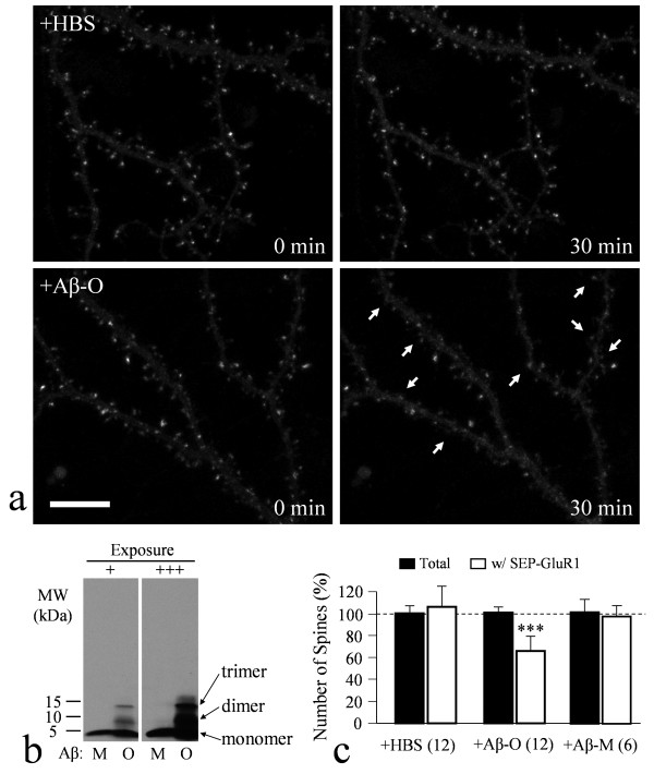
Figure 1.
Aβ-induced loss of surface AMPARs as revealed by confocal live-cell imaging of SEP-GluR1. (a) Representative images of dendritic regions of cultured hippocampal neurons (DIV21) expressing SEP-GluR1 before and after 30 min exposure to the control saline (HBS, upper panels) or 5 μM Aβ1-42 solution (lower panels). SEP-GluR1 signals are mostly seen concentrating in dendritic spines. Arrows indicate the spines exhibiting substantial loss of SEP-GluR1 signals after 30 min exposure to Aβ. Scale bar: 10 μm. (b) Western blots showing the existence of Aβ monomers and oligomers in our Aβ preparation. Two different exposures (+ short, +++ long) were shown to ensure that no additional Aβ aggregates in Aβ-M and Aβ-O preparations. (c) Quantitative analysis showing the number of total spines or spines exhibiting strong SEP-GluR1 fluorescence after 30 min exposure to HBS or Aβ. The data were normalized against the number before the 30 min exposure with 100% indicating no change. Triple asterisks: p < 0.0005 comparing to the corresponding control group (Student's t-test).

