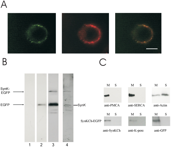
Figure 2. Expression of SynK in Chinese Hamster Ovary cells.
A) SynK-EGFP fusion protein expression in CHO cell plasma membrane, revealed by fluorescence microscopy. Fusion protein (left image) and PM-specific Vybrant DiI dye (central image) co-located as indicated by overlapping image (right). Representative images are shown. Bars: 10 µm. Unequal distribution of Vybrant DiI may be due to preferential concentration of dye in rafts or to rapid vesicular uptake. B) SynK-EGFP is expressed with predicted molecular weight in CHO cells. Untransfected cells (lane 1) and CHO cells transfected with pEGFP-N1 (lane 2) or pSynK-EGFP (lanes 3, 4) were lysed 72 h after transfection, and 50 µg (lanes 1, 2, 4) or 100 µg (lanes 3) total proteins were loaded. Membranes were developed with anti-GFP (lanes 1–3) or anti-SynK (lane 4) primary antibodies. Arrows: positions of EGFP (28 kDa), SynK (27 kDa) and SynK-EGFP (54 kDa) proteins. C) SynK fusion protein is revealed in membraneous fraction. The purity of soluble and membrane fractions obtained from transfected CHO cells was checked by antibodies against marker proteins of the plasmamembrane (PMCA) (140 kDa), endoplasmatic reticulum (SERCA) (110 kDa) and cytosol (actin) (42 kDa) (upper panels). Actin is found also in the membraneous fraction because it is in part associated to organelles and cytoskeletron. SynK-EGFP fusion protein is present in the membraneous fraction (lower panels). Equal volumes of pellet and supernatant fractions, obtained as described in the Material and Method section, were loaded on SDS-PAGE (25 µl for samples developed with anti-SynK and anti-KPORE and 15 µl for those developed with anti-GFP antibody).

