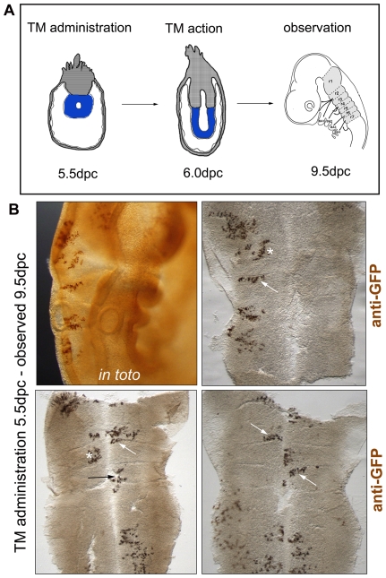
Figure 1. Clones respect the rhombomeric boundaries upon early labelling.
(A) Scheme depicting the embryonic stage upon TM administration (5.5 dpc), the embryonic stage of Cre-induction (6.0 dpc), and the stage when the observation was carried out and the hindbrain dissected (9.5 dpc). Note the dramatic change in embryo morphology between the time of TM administration and the time of embryo observation. Blue areas in 5.5–6.5 dpc depict the embryonic tissue. r1-r7, rhombomere 1–7. (B) Immunostaining to reveal GFP-positive clones in whole embryos (in toto) and in flat-mounted hindbrains.

