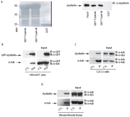
Figure 3. Dysferlin complexes with alpha-tubulin.
A. GST, GST-TubA4A and GST-TubA1B fusion proteins immobilized onto glutathione-Sepharose 4B beads were incubated with mouse skeletal muscle homogenate. GST, GST-TubA4A or GST-TubA1B with adsorbed proteins from mouse skeletal muscle were resolved by SDS-PAGE, transferred onto a nitrocellulose membrane, and blotted with mouse monoclonal anti-dysferlin antibody. Left panel: nitrocellulose membrane stained with ponceau red, right panel: detection of immunoreactive dysferlin. B. GFP-dysferlin was overexpressed in HEK293T cells and then immunoprecipitated from cell extracts with anti-GFP antibody. As a control, protein A-Sepharose beads coated with anti-GFP antibody were incubated with extracts of non-transfected HEK293T cells. Proteins were separated on SDS-PAGE gel and were transferred onto a nitrocellulose membrane and blotted with anti-GFP or anti-alpha-tubulin antibodies. Input (right panel), immunoprecipitate (left panel). C–D. Co-immunoprecipitation of dysferlin with anti-alpha-tubulin antibody from C2C12 myotube extracts (C) or mouse skeletal muscle homogenate (D). Input (right panel), immunoprecipitate (left panel). As a control (CTL), protein A-Sepharose beads were incubated with myotube extracts in the absence of anti-alpha-tubulin antibody.

