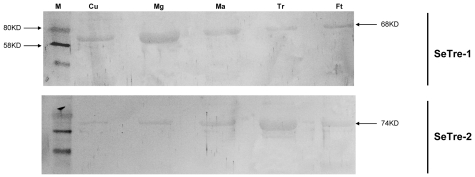
Figure 2. Immuno-blot analysis of SeTre-1 and SeTre-2 in various tissues.
10 ug protein extracts from the crude tissue extracts were loaded on 12 % SDS-PAGE. Lanes were immuno stained with anti-SeTre-1 serum or anti-SeTre-2 serum. Arrows indicated the positions of molecular weight markers. Cu: cuticle; Mg: midgut; Ma: Malpighian tubules; Tr: tracheae; Ft: fat body.

