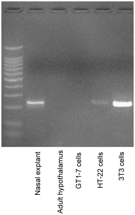
Figure 1. FGF expression in mouse brain and representative cell lines.
Photomicrograph of RT-PCR product for FGF8 mRNA stained with ethidium bromide and resolved on a 2% agarose gel. Total RNA was isolated from mouse nasal explant (embryonic day 11.5; lane 2) adult hypothalamus (lane 3), hypothalamic-derived GT1-7 cells (lane 4), hippocampus-derived HT-22 cells (lane 5), and fibroblast-derived 3T3 cells (lane 6). Presence of band indicates FGF8 primary transcripts in representative tissue type or cell line.

