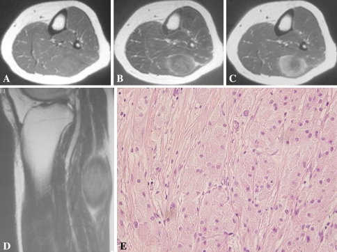
Fig. 1A–E.
The imaging and histologic characteristics of a representative case of benign granular cell tumor are shown. The patient is a 36-year-old man (Patient 4) with a tumor located in the lateral head of the gastrocnemius. (A) An axial T1-weighted MR image shows the tumor with a signal isointense to skeletal muscle. The signal intensity is slightly heterogeneous. (B) An axial T2-weighted image shows the tumor with slightly higher-intensity signal than the skeletal muscle and a heterogeneous signal with ill-defined margins with a peripheral zone of edema. (C) An axial T1-weighted image after gadolinium administration shows the tumor with diffuse enhancement. (D) A sagittal T2-weighted image of the knee shows the tumor with a peripheral heterogeneous high-intensity signal. (E) A histologic specimen shows characteristic sheets of polygonal tumor cells (Stain, hematoxylin and eosin; original magnification, ×400).

