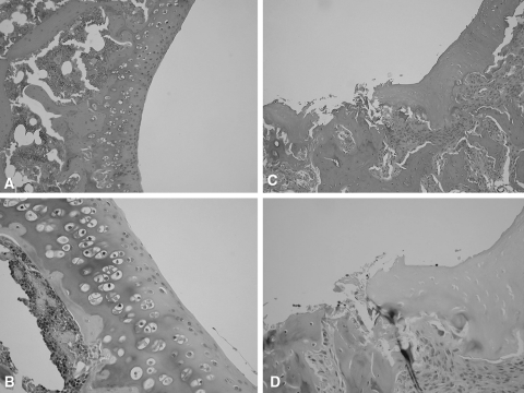
Fig. 3A–D.
(A) No abnormality is present in this normal rat knee (Stain, hematoxylin and eosin; original magnification, ×10), (B) or this normal rat knee (Stain, Safranin-O; original magnification, ×20). (C) The irregular surface of the joint cartilage and exposure of subchondral bone are evident in this monosodium iodoacetate (MIA) rat knee (Stain, hematoxylin and eosin; original magnification, ×10). (D) Ulceration and fibrillation of the cartilage with inflammatory cell infiltration are present in this MIA rat knee (Stain, Safranin-O; original magnification, ×20).

