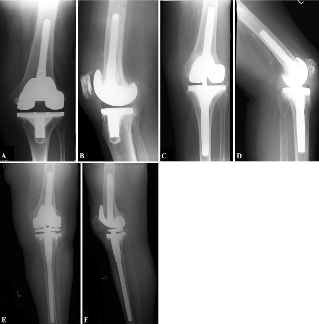
Fig. 1A–F.
Radiographic images of a modular posterior stabilized implant are shown from (A) the anteroposterior view and (B) the lateral view. Radiographic images of a condylar constrained knee implant are shown from (C) the anterioposterior view and (D) the lateral view. Radiographic images of a rotating hinge implant are shown from (E) the anteroposterior view and (F) the lateral view.

