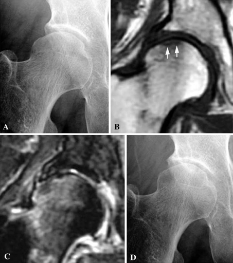
Fig. 1A–D.
A 76-year-old woman was treated nonoperatively for SIF. (A) Her initial radiograph shows a slight collapse of the lateral portion of the right femoral head. (B) A coronal T1-weighted fast spin echo MR image shows a low signal intensity band in the subchondral area (white arrows) associated with diffuse low signal intensity in the femoral head and neck. (C) A coronal fat-suppressed T2-weighted fast spin echo MR image shows a linear pattern of low signal intensity in the subchondral area with diffuse high signal intensity in the femoral head and neck. (D) A radiograph taken 10 months after the onset of hip pain shows no obvious change. The patient has only slight hip pain when walking.

