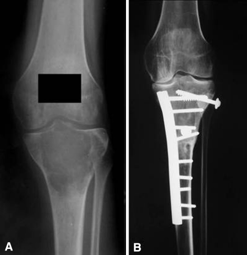
Fig. 1A–B.
The radiographs illustrate the case of a 25-year-old woman with a diagnosis of giant cell tumor who had a proximal tibia osteoarticular allograft after resection of the tumor (Case 37). (A) A preoperative AP radiograph shows the extension of the tumor that compromises the proximal end of the tibia. (B) The 8-year radiographic control shows a solid union of the osteotomy.

