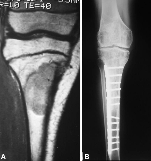
Fig. 2A–B.
The images illustrate the case of a 12-year-old girl with a diagnosis of Ewing’s sarcoma who received chemotherapy and had an osteoarticular allograft after tumor resection of the proximal tibia (Cases 13 and 15). Owing to a deep infection, the osteoarticular allograft was removed (Case 13) and antibiotic-impregnated polymethylmethacrylate spacer cement was placed. After achieving infection control, a second osteoarticular allograft was performed (Case 15). (A) A coronal MR image obtained after chemotherapy but before the first surgery was performed shows the tumor extension. (B) An AP radiograph of the second osteoarticular allograft after 16 years of followup shows joint deterioration.

