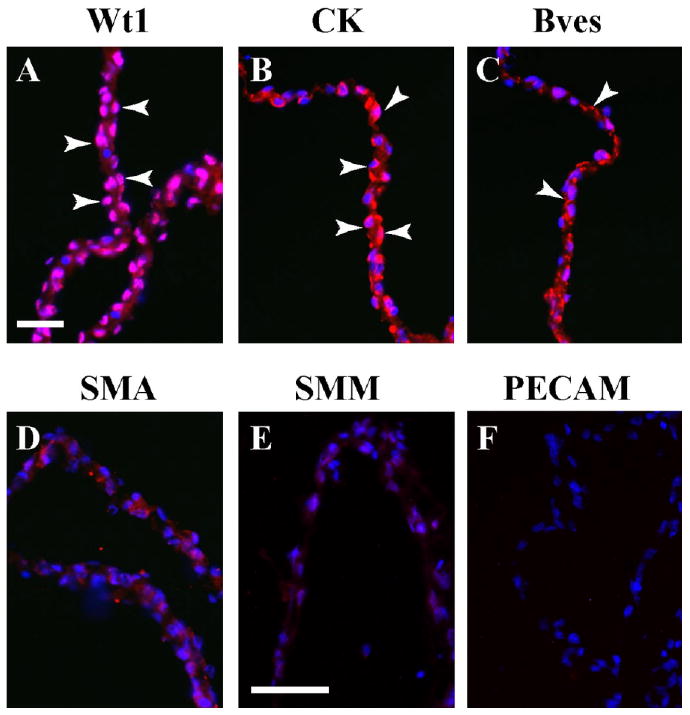
Fig. 1.

Characterization of serosal mesothelium using marker proteins. Freshly excised serosal mesothelium preparation was sectioned and stained with specific marker antibodies. A,B: This preparation reveals nuclear Wt1 expression (A, arrowheads) and cytokeratin (CK) distribution (B, arrowheads). C–F: Bves is found at cell–cell contacts (C, arrowheads), while SMA (D), SMM (E), and PECAM (F) are not detectable above background. Scale bars = 50 μm in A–F.
