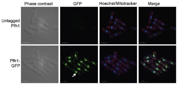
Figure 5.
SpPfh1 is detected in both the nucleus and mitochondria. Wild type cells (untagged Pfh1) and cells expressing Pfh1 fused with GFP at the C-terminus (Pfh1-GFP) are viewed by phase contrast and fluorescence microscopy. Pfh1 is visualized by GFP (green), DNA by Hoechst (blue), and mitochondria by mitotracker (red). The white arrow points out concentrated Pfh1-GFP in the nucleolus. The scale bar indicates 10 μm. This figure is adapted from the journal of Molecular and Cellular Biology, copyright © American Society for Microbiology [Molecular and Cellular Biology , Vol 28, 2008, p.6598, doi:10.1128/MCB.00191-08] [72].

