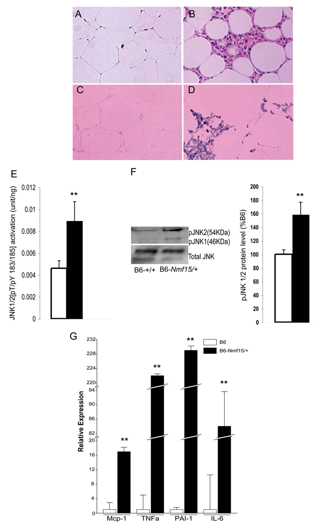
Figure 7. Macrophage infiltration in adipose tissue of obese B6-Nmf15/+ mice at 25 weeks of age.
H.E. staining of adipose tissue (A) and (B). F4/80-positive macrophages were detected with a F4/80 antisense riboprobe (C) and (D). Increased JKN1/2 phosphorylation in inflammatory adipose tissue (E and F). Adipose tissue cytokine gene expression by real time PCR (G). Representative images from a total of three mice examined are shown. *, p < 0.05, **, p<0.01 for B6-Nmf15/+ samples versus the respective B6-+/+ samples.

