Figure 2.
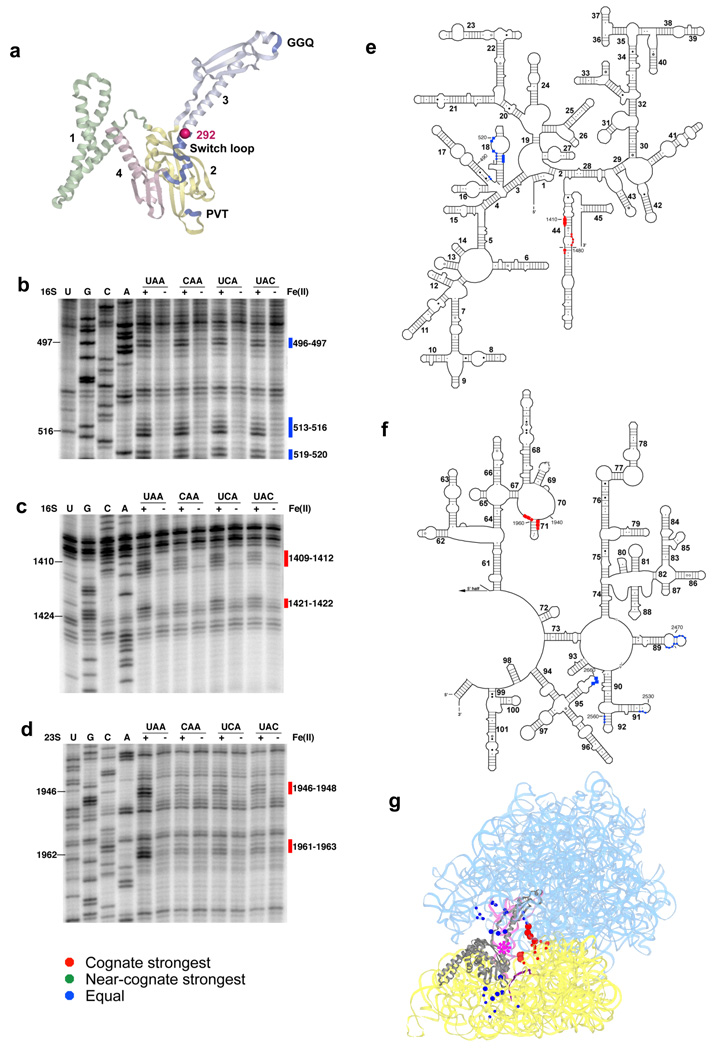
Directed hydroxyl radical probing of the ribosome environment of the switch loop of RF1 in ribosome complexes programmed with various codons in the A site. (a) Ribbon diagram of RF1. Sphere indicates Cα of residue 292 where Fe(II) is tethered. (b–d) Primer extension analysis of cleavage of 16S rRNA (b–c) and 23S rRNA (d) from saturating levels of Fe(II)-S292C-RF1 incubated with the indicated ribosome complexes with primers (b) 565 (c) 1490 and (d) 2042. (e–f) Directed hydroxyl radical cleavage sites from Fe(II)-S292C-RF1 shown on secondary structure of (e) 16S rRNA and (f) the 3′ half of 23S rRNA. (g) All cleavage sites modeled on tertiary structure of RF1 bound ribosome complex (PDB entry: 3D5A10 and 3D5B10), as in Figure 1.

