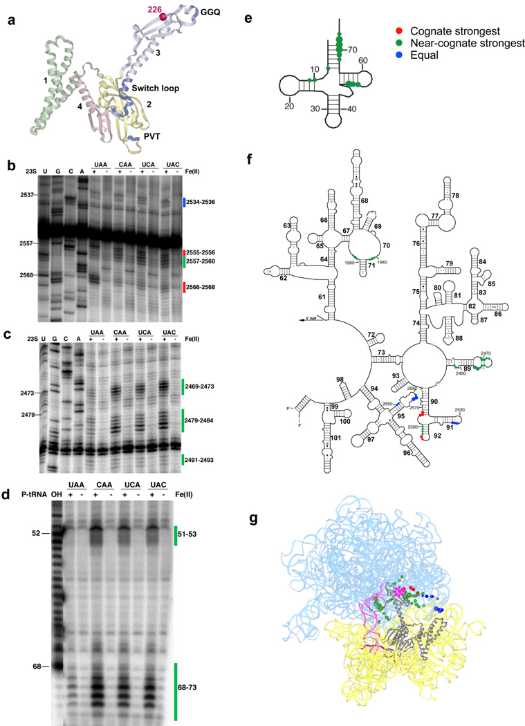
Figure 3.
Directed hydroxyl radical probing of the ribosome environment of the catalytic motif “GGQ” of RF1 in ribosome complexes programmed with various codons in the A site. (a) Ribbon diagram of RF1. Sphere indicates Cαof residue 226 where Fe(II) is tethered. (b–c) Primer extension analysis of 23S rRNA from saturating levels of Fe(II)-T226C-RF1 incubated with the indicated ribosome complexes with primers (b) 2639 and (c) 2542. (d) Denaturing sequencing gel analysis of cleavage of P-site tRNA from Fe(II)-T226C-RF1. (T1) cleavage by RNaseT1; (OH) alkaline hydrolysis ladder. (e–f) Directed hydroxyl radical cleavage sites from Fe(II)-T226C-RF1 shown on secondary structure of (e) P-site tRNA and (f) the 3′ half of 23S rRNA. (g) All cleavage sites modeled on tertiary structure of RF1 bound ribosome complex (PDB entry: 3D5A10 and 3D5B10), as in Figure 1.

