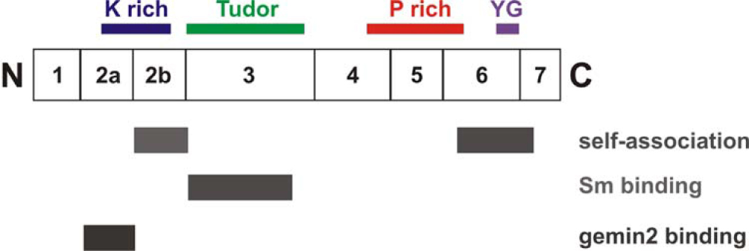
Figure 2B. Domains of SMN.
A diagram of SMN showing the exons and domains. Exon 2B encodes a domain important in binding Gemin2 as well as self-association.160 Both exon 2A and 2B are conserved. The K domain is rich in lysine, the Tudor domain is in exon 3 and has homology to other Tudor domains. The Tudor domain binds Sm proteins.97 Exon 5 and part of exon 6 contain a proline rich domain that may influence profilin binding.161 The C-terminal domain of exon 6 contains the conserved YG box and is important for self-association.160, 162

