
Figure 5. Mtp, Ire1β and Nr2f1 expression in mouse intestine.
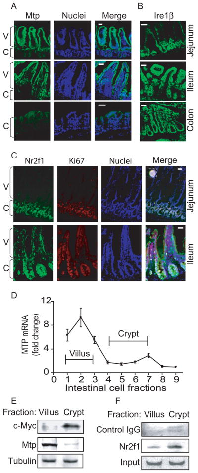
Frozen mouse proximal jejunum, distal ileum, and colon were sectioned (10 μm) and processed for staining as in Figure 4B.
(A–B) Goat anti-MTP (sc-331166) (A) and IRE1β (sc-10512) (B) were incubated (1h) with sections. Anti-goat Alexa Flour 488 antibody was added and nuclei were stained with Topro3 blue. Bar, 100 μm; C, crypt; V, villus
(C) Goat anti-NR2F1 and rabbit anti-Ki67 (Vector Labs, Burlingame, CA) antibodies were used for staining the intestinal sections.
(D–F) Mouse jejunum (n=3, 16 cm from duodenum) was incubated with 1% EDTA for various times to separate villus and crypt cells from the intestinal wall as described in Supplementary Materials and Methods. Mtp mRNA was determined with qRT-PCR (D). Cells representing villi (fractions 1–3) and crypts (fractions 4–7) were pooled and presence of c-Myc, Mtp, and tubulin was determined by western blotting (E). Binding of NR2F1 to the MTTP promoter in villus and crypt cells was analyzed by ChIP followed by regular PCR. Goat serum IgGs were used as a negative control (F).