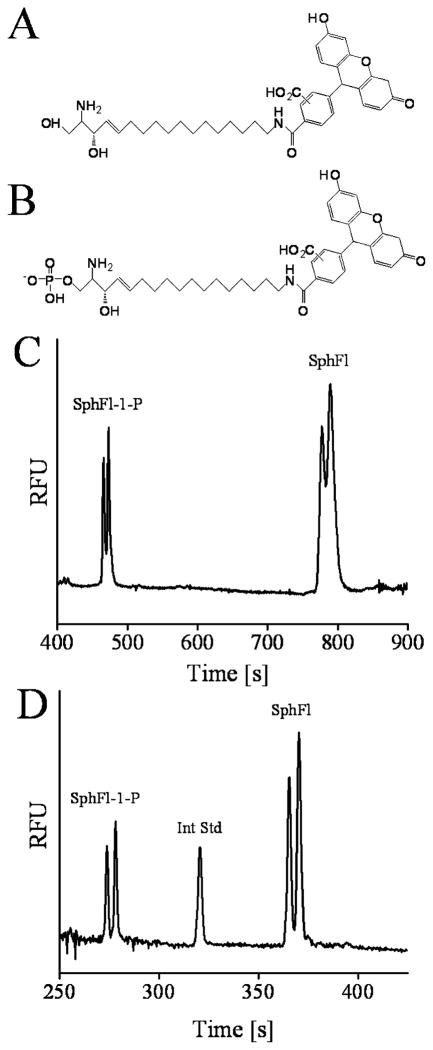
Figure 1.
Chemical structures of (A) fluorescein-labeled sphingosine (SphFl) and (B) fluorescein-labeled sphingosine 1-phosphate (SphFl-1-P). (C) Electropherogram of the separation of SphFl-1-P (7.2 × 10⁻⁹ M) and SphFl (1.4 × 10⁻⁸ M) in a nonmicellar buffer (100 mM Tris, 20% 1-propanol, 5% EOTrol LR at pH 8.5). (D) Separation of SphFl-1-P (4.3 × 10⁻⁹ M) and SphFl (8.5 × 10⁻⁹ M) including an internal standard (Int Std) of Bodipy-Fl (5.1 × 10⁻⁹ M) with the addition of 10 mM SDC to the electrophoresis buffer.

