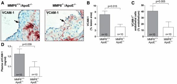
Figure 6. Reduced VCAM-1 expression in MMP8−/−/apoE−/− mice compared with MMP8+/+/apoE−/− mice.
Panels A, B and C show results of immunohistochemical analyses of VCAM-1 in aortic atherosclerotic lesions, (A), representative images; (B), mean VCMA-1 staining in lesions; (C), mean VCAM-1 staining in the endothelial layer. Panel D shows mean levels of soluble VCAM-1 in plasma. Error bars in the column charts are standard error of mean.

