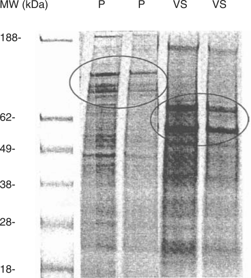
Figure 1.
SDS-polyacrylamide electrophoresis (SDS-PAGE) of seminal prostasomes (P) and vesicular seminalis secretion (VS) at two different concentrations. Molecular markers in lane 1 (MW). Prostasome marking CD proteins (at 150, 120 and 90 kDa) encircled in the SDS-PAGE pattern lanes marked P. HSP70 and clusterin (at 70 and 55 kDa) encircled in SDS-PAGE pattern lanes marked VS.

