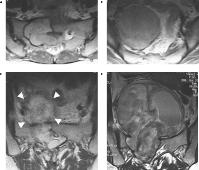
Figure 4.
Axial image of T2-weighted MRI showing a big tumor extending from the spinal canal through the sacral body of S1 (A) to the presacral region (S2 level (B)) before surgery. The tumor was partially removed via a combination of the anterior and posterior approach. Axial image of T2-weighted MRI immediately after the surgery (C) showing a postoperative hematoma in the remaining tumor capsule (white arrow-head). Seven years after the surgery, the remaining tumor grew (D), and complete removal of the tumor with S1 root sacrifice was performed.

