
Figure 1.
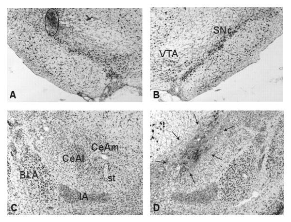
Photomicrographs showing representative brain sections of SNc and CeA of an animal that received a cannula implant targeting SNc in the left hemisphere and a CeA lesion in the right hemisphere. A, B Nissl-stained sections of the left and right SNc, respectively. Open circle shows mild gliosis caused by the tip of injection cannula. C, D, Nissl-stained sections of the intact and lesioned (arrows) CeA, respectively. BLA, basolateral amygdala; CeAl, lateral CeA; CeAm, medial CeA; IA, intercalated nucleus of the amygdala; st, stria terminalis (encircled by dotted line); VTA, ventral tegmental area.