Figure 1.
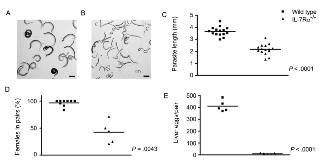
Schistosoma mansoni development in interleukin-7 receptor α–deficient (IL-7Rα−/−) mice. IL-7Rα−/− and wild-type mice were infected percutaneously with S. mansoni cercariae, and parasite development and egg production were assessed at 6 weeks after infection. A, S. mansoni worms isolated from wild-type mice. B, S. mansoni worms isolated from IL-7Rα−/− mice. C, Length of male S. mansoni worms isolated from wild-type and IL-7Rα−/− mice. D, Percentage of female worms participating in pairs in wild-type and IL-7Rα−/− mice. E, No. of eggs deposited per pair of worms in the livers of wild-type and IL-7Rα−/− mice. The scale bars in panels A and B represent a length of 1 mm. P values were calculated using the Mann-Whitney U test.

