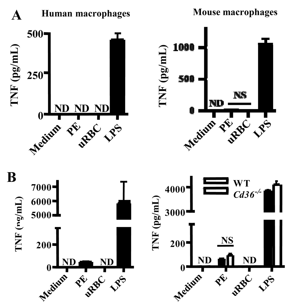
Fig. 3. CD36-mediated internalization of P. falciparum PEs by macrophages does not stimulate pro-inflammatory cytokine secretion.
(A) Human macrophages (left) and wild-type murine macrophages (right) were incubated for 4 hr at 37°C with carefully synchronized and washed mature-stage PEs, uninfected red blood cells (uRBC), or LPS. Supernatants were collected and analyzed by ELISA for TNF. NS, not significant by Student’s t-test. (B) To ensure that lack of priming was not obscuring inflammatory consequences of CD36-mediated PE internalization, macrophages were pre-incubated for 12 hr with IFN-γ (100 U/mL) prior to the internalization assay. Cd36−/− murine macrophages were included as a control to assess the contribution of CD36-mediated PE internalization to the low levels of TNF production observed. NS, not significant by Student’s t-test (human data); *** indicates p<0.001 and NS indicates not significant by one-way ANOVA with Bonferroni post-tests (murine data). ND, not detectable. Results are representative of three independent experiments.

