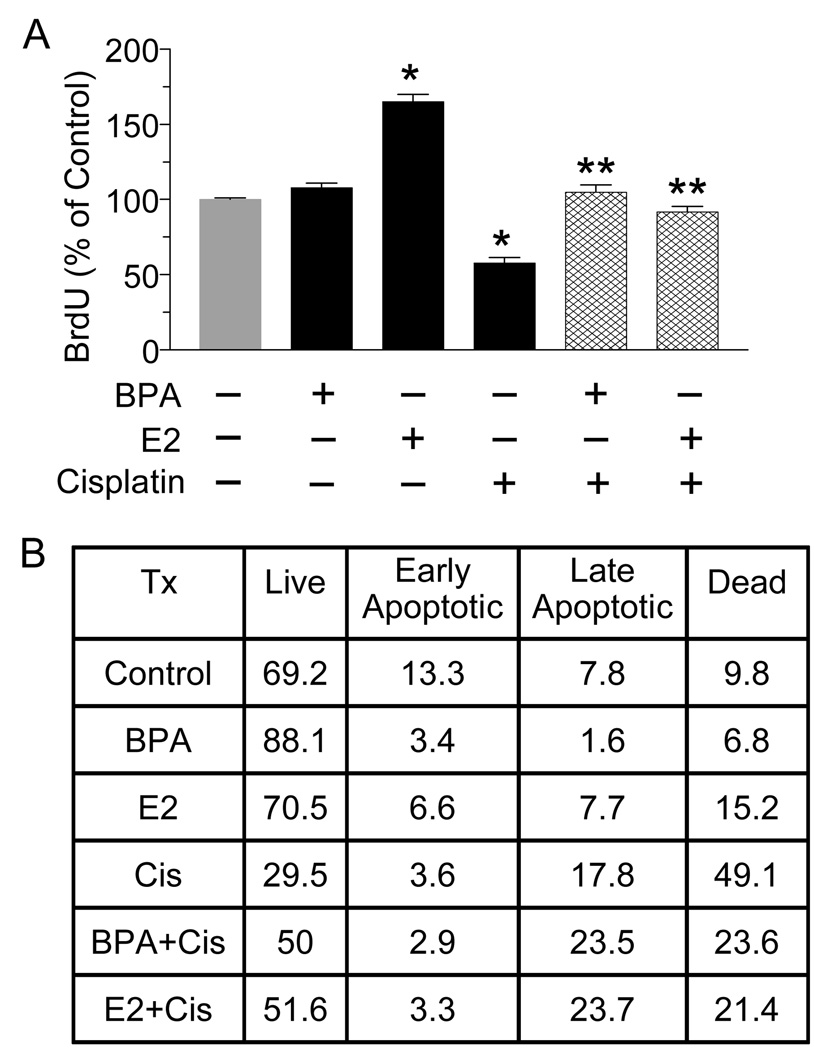
Fig. 2.
BPA and E2 protect T47D cells from cisplatin-induced decreases in cell proliferation and increases in apoptosis. (A) T47D cells were treated with 10 nM BPA or E2 for 24 hrs followed by cisplatin (800 ng/ml) for 72 hrs. Cells were fixed and incubated with a BrdU antibody for 8 hrs. After adding substrate, the product was quantified by measuring absorbance. Each value is a mean±SEM of four replicates. * significant (p< 0.05) vs control. ** significant vs cisplatin. (B) T47D cells were treated as in (A), stained with Annexin V and propidium iodide (PI), and analyzed by flow cytommetry. The table depicts the percentage of cells in each treatment group that are alive (no stain), in early apoptosis (Annexin V-positive), late apoptosis/necrosis (Annexin V+PI-positive) or dead (PI- positive). Shown is a representative experiment repeated 3 times

