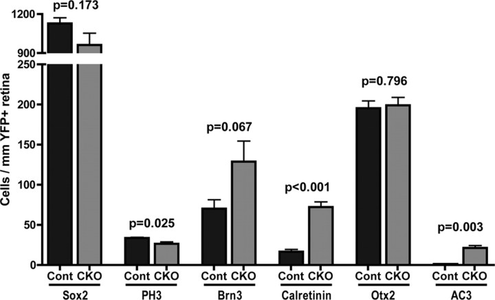
Figure 2.
Quantification of immunofluorescence staining of E16 control and Dicer CKO retinas. Counts of Sox2+ retinal progenitor cells, PH3+ M-phase cells, Brn3+ ganglion cells, calretinin+ horizontal cells, Otx2+ photoreceptors, and apoptotic cells (AC3+) in the neuroblastic layer per millimeter of YFP+ retina. Error bars indicate mean ± SEM.

