
Figure 3.
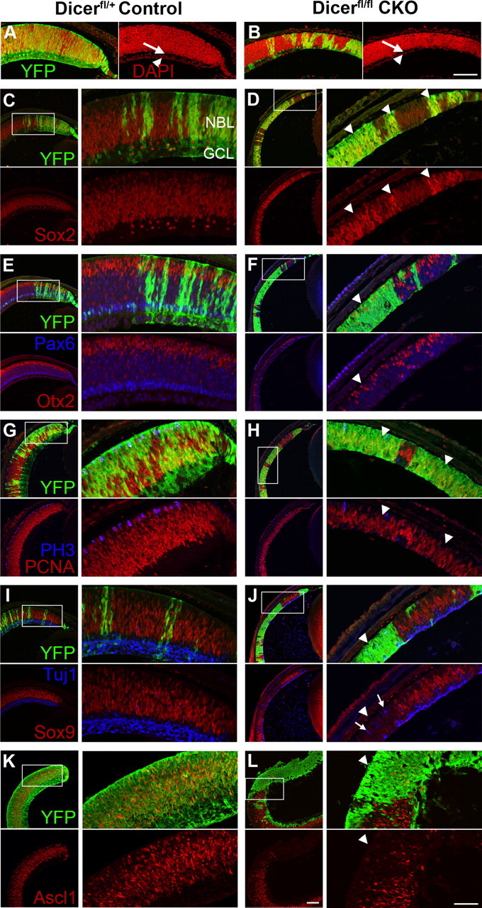
Immunofluorescence staining of early postnatal control and Dicer CKO retinas. A–J, P0/P1 retinal paraffin sections. YFP staining (green) indicates areas of cre-mediated recombination and Dicer CKO. K, L, P0 + 2 DIV retinal cryosections. YFP staining in green. A, B, Retinal lamination is affected in Dicer CKO retinas, with an absent ganglion cell layer (arrowhead) and inner plexiform layer (arrow) as revealed by nuclear DAPI (red) staining. C, D, Sox2 RPC staining (red) is increased in Dicer-deficient areas (arrowheads). E, F, Otx2+ photoreceptors (red) are decreased in Dicer-deficient areas (arrowhead). Bright Pax6+ amacrine cells (blue) are absent from Dicer-deficient areas, although fainter RPC Pax6 staining appears normal (arrowhead). G, H, Dicer-deficient areas (arrowheads) show reduced PCNA staining (red) and have fewer PH3+ cells (blue). I, J, Dicer-deficient areas (arrowhead) lack Sox9+ RPCs (red) and have ectopic Tuj1+ neurons (blue, arrows). K, L, Dicer-deficient areas (arrowhead) lack Ascl1+ RPCs (red). NBL, Neuroblastic layer; GCL, ganglion cell layer. All images are oriented the same, with the ganglion cell layer on the bottom and peripheral retina to the right. Boxes represent areas of magnification. Scale bars: 200× images, 100 μm; 400× insets, 50 μm.