
FIGURE 1.
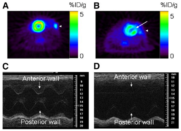
Cardiac functional assessment using 18F-FDG PET and high-resolution ultrasound (at frequency of 30 MHz). (Top) 18F-FDG PET of sham-operated animal (A) and animal after MI (B). Sham operation did not induce any 18F-FDG defect, whereas MI was associated with medium-sized defect in anterolateral wall (white arrow). (Bottom) M-mode ultrasound at midventricle level in sham-operated animal (C) and animal after MI (D). After MI, there was akinesis of anterolateral wall and significant decrease in fractional shortening compared with that of sham-operated animals.