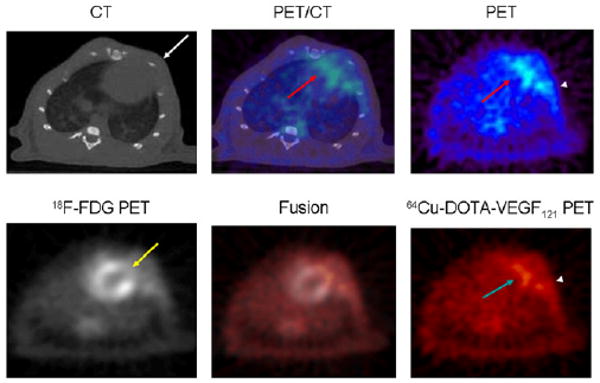
FIGURE 2.

Myocardial origin of 64Cu-DOTA-VEGF121 PET signal after MI. (Top) Representative coregistered images of microCT (left), PET (right), and fused PET/CT image (center) in MI animal clearly demonstrates that the 64Cu-DOTA-VEGF121 signal detected with PET corresponds to anterolateral myocardium (PET and fused images, red arrow) and clearly separated from intercostal muscle layer (microCT image, white arrow). There is also increased uptake in area of surgical wound (PET image, arrowhead). (Bottom) Representative images of 64Cu-DOTA-VEGF121 (left), 18F-FDG (right), and 64Cu-DOTA-VEGF121/18F-FDG fused image (middle). 18F-FDG scan shows that coronary artery ligation resulted in lack of 18F-FDG uptake (yellow arrow) and that uptake of 64Cu-DOTA-VEGF121 occurs in areas supplied by ligated coronary artery (turquoise arrow). Fusion of both scans results in complementation of 18F-FDG and 64Cu-DOTA-VEGF121 signals. There is also increased uptake in area of surgical wound (arrowhead).
