FIGURE 3.

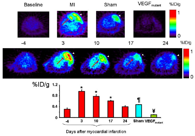
(Top) Representative images at baseline (left), animals after MI (middle left), and sham-operated animals (middle right) show difference in myocardial uptake in MI animals compared with baseline and sham animals. Red arrow shows the 64Cu-DOTA-VEGF121 signal from myocardium (seen only in MI animals), and arrowheads show 64Cu-DOTA-VEGF121 signal from the surgical wound (muscle layer), which is present in both sham-operated and MI animals. Right panel shows typical image acquired using VEGFmutant, with minimal uptake, supporting the specificity of 64Cu-DOTA-VEGF121 probe for VEGFRs. (Middle) Representative images correspond to 1 animal of MI group illustrating uptake of 64Cu-DOTA-VEGF121 over time (in days after induction of MI), clearly showing time-dependent effect on uptake of 64Cu-DOTA-VEGF121. Red arrow points to myocardial upake, whereas white arrowhead points to chest wall muscular layer uptake. (Bottom) Quantification of 64Cu-DOTA-VEGF121 after MI over time, expressed in %ID/g of tissue. 64Cu-DOTA-VEGF121 uptake was highest on day 3 postoperatively (compared with baseline, day −4) and continues to be elevated until day 17 postoperatively. 64Cu-DOTA-VEGF121 uptake was also significantly different compared with sham-operated animals and VEGFmutant. *P < 0.05 compared with baseline; ¥P < 0.05 compared with sham and 64Cu-DOTA-VEGF121; ¶P < 0.05 compared with VEGFmutant and 64Cu-DOTA-VEGF121.
