
FIGURE 4.
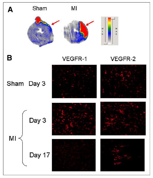
Ex vivo studies. (A) Autoradiography of 30-μm myocardial slices of both sham-operated (left) and MI (right) animals after injection of 64Cu-DOTA-VEGF121 shows increased signal in anterolateral wall of LV of MI animals, whereas no activity is detected in sham group. Red arrows point to area affected by ligated artery (anterolateral wall), clearly showing the myocardial origin of signal observed. (B) Immunofluorescence staining for VEGFR-1 (left) and VEGFR-2 (right) in sham-operated and MI animals (on days 3 and 17 after MI). MI is associated with marked increase in VEGFR-1 and VEGFR-2 immunostaining, which was higher than that of sham animals. VEGFR expression is higher on day 3 and diminishes over time, similar to what it is observed with PET. MIN = minimum; MAX = maximum.