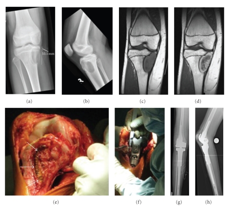
Figure 1.
Pre- and postoperative radiological imaging of the right leg and in situ pictures: (a and b) preoperative conventional X-rays of the right knee. The tumor shows an eccentric osteolytic lesion located at the epimetaphyseal proximal tibia, not respecting the epiphyseal plate. The medial cortical bone is completely destroyed and the formation of partially calcified periosteal lining suggests a Lodwick-type 1C lesion. (c) MRI scan demonstrating a T1 isointense, T2 hypointense tumor formation. A similar zonal architecture with a large central core of very low signal intensity and a peripheral rim of intermediate to high signal intensity on T1- and T2-weighted spin-echo pulse sequences was observed by Christensen et al. [30]. (d) MRI scan showing high uptake of Gadolinium predominantly at the tumor's periphery. A thin layer of sclerotic bone separates the tumor from circumjacent marrow edema. (e and f) Intraoperative images during tumor resection and implantation of the endoprosthesis. Resection of the tumor was performed in no-touch technique. The scar of the previous open biopsy (white arrows) as well as the former access path remained on the resected bone. Reconstruction was accomplished by implanting a partially custom-made tumor endoprosthesis in combination with linked knee replacement. (g and h) Postoperative radiographs of the right leg show the endoprosthesis in proper position.

