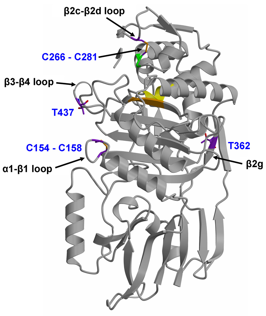
Fig. 3.
Location of two disulphide bridges, and two threonines proposed to be targets of phosphorylation by PknB 20, in PBPA. Set against the fold of PBPA (displayed in grey ribbon) are the cysteines that form disulphide bridges and threonines 362 and 437. In both cases, carbon bonds are colored purple. Nearby elements of secondary structure to each site are marked by arrows. The active site is indicated by the cluster of conserved motifs, with the ribbon colored yellow for the SxxK motif, green for the SxN motif and orange for the KTG motif.

