Figure 2.
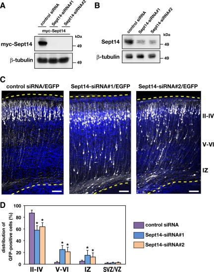
Roles of Sept14 in neuronal migration. (A) pCAG-myc-Sept14 was cotransfected into COS7 cells with control siRNA, Sept14-siRNA#1 or Sept14-siRNA#2. After 72 h, cells were lysed and subjected to immunoblotting with anti-myc or anti-β-tubulin. (B) Cortical neurons were transfected with control siRNA, Sept14-siRNA#1, or Sept14-siRNA#2. After 72 h, cells were lysed and subjected to immunoblotting with anti-Sept14 or anti-β-tubulin. (C) pCAG-EGFP was coelectroporated with control siRNA, Sept14-siRNA#1, or Sept14-siRNA#2 into cerebral cortices at E14.5 and fixed at P2. Coronal sections were immunostained with anti-GFP (white). Nuclei were stained with DAPI (blue). Dotted lines represent pial and ventricular surfaces. Bars, 100 μm. II–IV, layers II–IV of CP; V–VI, layers V and VI of CP. (D) Quantification of the distribution of EGFP-positive cells in distinct parts of the cerebral cortex (layer II–IV, layer V–VI, IZ, and SVZ/VZ) for each condition shown in C. Error bars indicate SD. *p < 0.01, percentage of cells in each region relative to the corresponding values with control condition.

