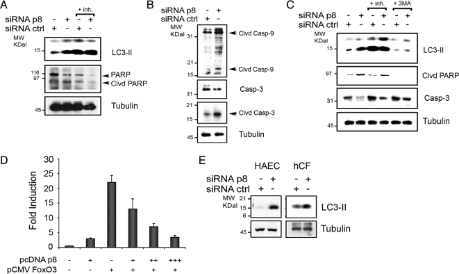
Figure 6.
p8 silencing in cardiovascular cells. (A–C) p8 silencing increases autophagy and apoptosis in cardiac cells. p8 RNAi in primary neonatal rat cardiomyocytes (A and B) and in rat H9C2 cardiac cells (C). Cells were cultured for 72 h in 10% FBS after (rat) p8 or ctrl RNAi. When indicated, both pepstatin A and E64D were added for 4 h (+inh) or 10 mM 3-methyladenine (+3MA) was added for 16 h before analysis. The Western blots were performed with anti-LC3B, PARP, rat-specific cleaved (clvd) caspase-9, and total and cleaved (clvd) caspase-3 antibodies for cardiomyocytes and LC3B and cleaved (clvd) PARP and caspase-3 antibodies for H9C2 cells. Tubulin was used as loading control. (D) p8 represses FoxO3 transactivation in a dose-dependent manner in H9C2 cells. H9C2 cells were transfected in triplicate with a FoxO luciferase reporter and pcDNAp8 and pCMVFoxO3 wt as described for Figure 3A). In separate experiments, FoxO3 was cotransfected with increasing amounts of pcDNAp8. Luciferase activity was measured in triplicate and normalized to coexpressed β-galactosidase activity. Means ± SEM for two experiments are shown. (E) p8 silencing increases autophagy in primary human cells. p8 RNAi in primary hCFs and in primary HAECs. Cells were cultured for 72 h in 10% FBS after p8 or ctrl RNAi, and the Western blot analysis was performed as described in A.

