Figure 3.
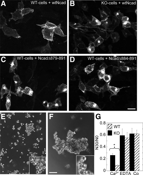
PTP1B expression and binding to N-cadherin cytoplasmic domain are required for N-cadherin targeting and function. WT-cells (A, C, and D) and KO-cells (B) transfected with GFP fusions of WT N-cadherin (A and B) or two deletion mutants which cannot bind PTP1B (C and D) were fixed and analyzed by fluorescence confocal microscopy. Note the similar phenotype between KO-cells (B) and WT-cells expressing the deletion mutants (C and D). Both display enhanced accumulation of fluorescence in Golgi and in scattered puncta. (E–G) Aggregation assays. Representative phase-contrast fields showing cell suspensions of KO-cells (E) and WT-cells (F) after 30-min incubation in HBSGK-1 mM CaCl2. Note that KO-cells form smaller and looser aggregates than WT-cells (E, F, and insets). Quantification of the aggregation in HBSGK alone (Co), or in the presence of 1 mM CaCl2 or 1 mM EDTA (see Materials and Methods) is shown in the graph (G). Data are means ± SEM of four experiments. Asterisk denotes significant difference (Student's t, p = 0.001). Bars, (A–D) 60 μm; (E and F) 150 μm.

