Figure 5.
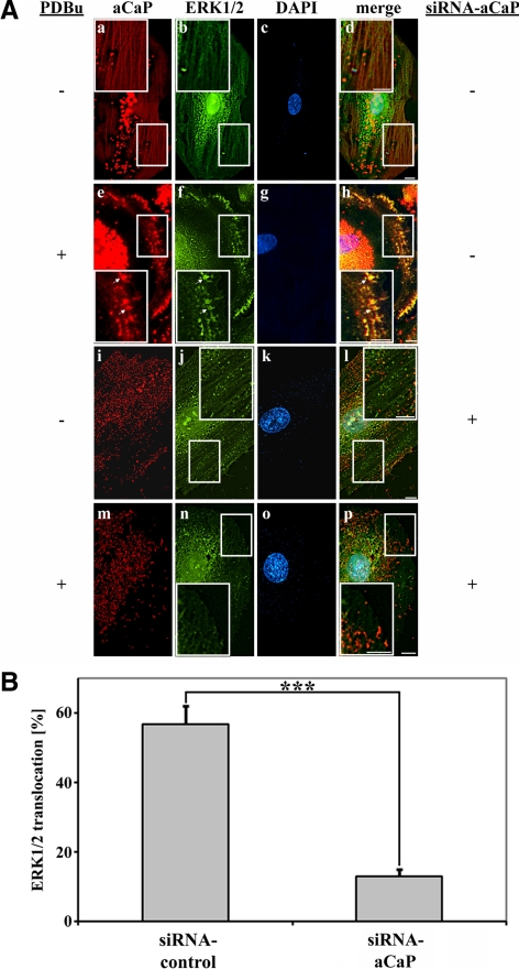
Knockdown of h3/acidic CaP inhibits PDBu-mediated ERK1/2 targeting. (A) REF52.2 cells were transfected with siRNA against aCaP siRNA (i–p) or nontargeting control siRNA (a–h). Three days after transfection, cells were treated with DMSO for control (a–d and i–l) or with 1 μM PDBu for 30 min (e–f and m–p) before staining for aCaP (a, e, i, and m) and ERK1/2 (b, f, j, and n) with the rabbit polyclonal anti-h3/acidic CaP-Al555 and anti-ERK1/2-Alexa488 antibodies. Note that the background in images a, d, e, h, i, l, m, and p derives from uncoupled Alexa555 dye, which was resistant to extensive washing. Images were obtained with deconvolution microscopy. The yellowish signal in the merged images indicates colocalization (d, h, l, and p). Bar, 10 μm. (B) REF52.2 cells transfected with siRNA against h3/acidic CaP or with nontargeting control siRNA were treated with 1 μM PDBu for 30 min. After an immunofluorescence staining for h3/acidic CaP and ERK1/2, cells showing an ERK1/2 translocation to either the cell cortex or podosome-like structures were counted. The graph represents three independent experiments, where at least 100 cells were counted. Note that the difference is highly statistically significant in a two-tailed paired t test (p = 0.0001) marked by asterisks.

